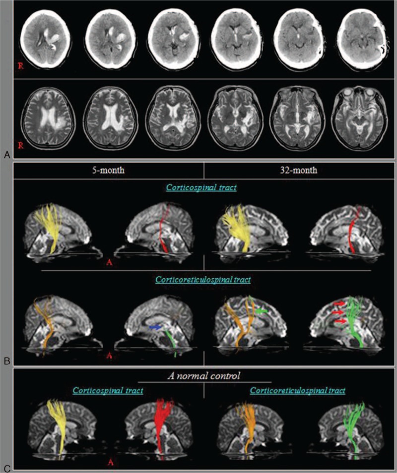
Figure 1.

A, Brain computed tomography (CT) images show hematomas in the left corona radiata and basal ganglia at onset and T2-weighted magnetic resonance (MR) images show a leukomalactic lesion in the left corona radiata and basal ganglia at 5 months after onset. B, Results of diffusion tensor tractography (DTT). The left corticospinal tract (CST) shows narrowing compared with the right CST on 5-month DTT. Similar findings were observed for both CSTs on 32-month DTT. The left corticoreticulospinal tract (CRT) shows discontinuation at the basal ganglia level on 5-month DTT (blue arrow) and this discontinuation is elongated to the cerebral cortex on 32-month DTT (red arrows), whereas on 32-month DTT, the right CRT has become thicker than it was on the 5-month DTT (green arrow). C, Results of DTT for the CST and CRT in a normal control.
