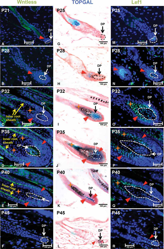
Figure 1. Wls is expressed in the DP during hair growth anagen except onset.
Mid-dorsal skin biopsies of wild-type C57BL/6 (B6) mice were collected at P21, P25, P28, P32, P35, P40 and P45, and processed for paraffin sections. Sections were analyzed for Wls (A–F) and LEF1 (M–R) expression by immunofluorescence staining at each time point as indicated. (G–L) Mid-dorsal skin biopsies of TOPGAL mice were collected at indicated age for X-gal whole-mount staining. The DP was circled by either white or black dash line in each hair follicle. Red triangles indicate either Wls-positive or Lef1-positive cells outside the DP in their respective pictures. Orange stars denote keratinocytes that are Lef1-positive but Wls-negative. Yellow arrows indicate inner root sheath cells in the hair follicle. For every indicated mouse age, at least three mice were analyzed. Scale bar: 100 µm.

