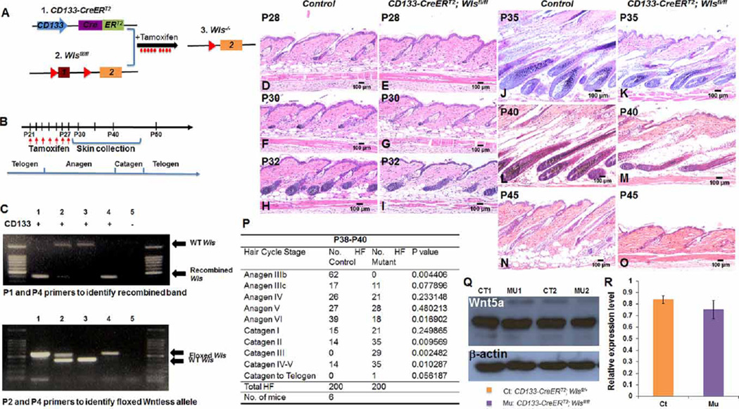
Figure 2. Ablation of Wls in CD133+ DP cells delays hair growth and induces premature hair regression.
(A) Illustration of the CD133-CreERT2; Wlsfl/fl transgenic mouse model, which allows specific Wls ablation in CD133+ DP cells upon TAM or 4-OHT administration. (B) Time scheme for TAM administration during early anagen stage and skin biopsy collection. CD133-CreERT2; Wlsfl/fl mice and control littermates were administered of tamoxifen daily by IP injection for 7 days starting from P21 to P27. Mid-dorsal skin biopsies were harvested for examination at P28, P30, P32, P35, P40 and P45. (C) Recombination of floxed Wls alleles in CD133+ DP cells was confirmed by PCR analysis. Upper DNA agarose gel picture: genomic DNA extracted from skin biopsies was PCR genotyped using P1/P4 primer set to identify wild-type Wls allele and recombined Wls allele. Lower DNA agarose gel picture: genomic DNA extracted from skin biopsies was PCR genotyped using P2/P4 primer set to identify wild-type Wls allele and floxed Wls allele. Lanes labeled with same number in upper and lower DNA gel pictures were genotyping results of genomic DNA extract from same mouse. Genotype of CD133-CreERT2 for each mouse is labeled above each lane of top DNA agarose gel picture. ‘+’ means mouse carried CD133-CreERT2 transgene, while ‘−’ means mouse did not carry CD133-CreERT2. (D–O) H&E-stained skin sections from CD133-CreERT2; Wlsfl/fl (D, F, H, J, L and N) and control littermates (E, G, I, K, M and O) as labeled. Scale bar: 100 µm (n = 3). (P) Comparison of hair follicle numbers that were present at different hair cycle stages between control and CD133-CreERT2; Wlsfl/fl mice from P38 to P40. A minimum of three skin biopsies from three pairs of mutant mice and control littermates were manually counted. Two-tailed paired Student’s t-test was employed. *P < 0.05. (Q) Generation of Wnt5a protein in CD133+ DP cells was confirmed by Western blot analysis. Two pairs of CD133-CreERT2; Wlsfl/fl mice (MU1, 2) and control littermates (CT1, 2) are shown. (R) Intensity of Wnt5a band was shown by scanning X-ray film and normalized to β-actin band. Three pairs of mice were analyzed.

