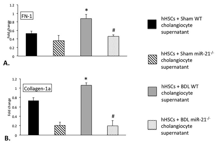
Figure 9.
Determination of hHSC fibrotic reaction, in vitro. hHSCs treated with supernatants extracted from cholangiocytes isolated from BDL WT mice show increased FN-1, and Collagen-1a expression when compared to hHSCs treated with supernatants from Sham WT mice (A, B); however, these parameters were decreased in hHSCs treated with cholangiocyte supernatants from BDL miR-21−/− mice when compared to BDL WT mice (A, B). Data are expressed as means ± SEM. n = 6 reactions from 12 sets of cells for qPCR. *p<0.05 versus hHSCs + WT cholangiocyte supernatants; #p<0.05 versus hHSCs + BDL WT cholangiocyte supernatants.

