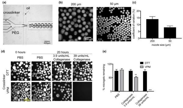
Fig. 1. Generation of protease degradable microgels using flow focusing microfluidics.
(a) Image of microfludic flow focusing device with 200 μm nozzle. (b) Image of microgels generated using a 200 μm (left) or 50 μm (right) nozzle. (c) Coefficient of variation of diameter for microgels generated from 200 μm or 50 μm nozzles. (d) Images of microgels crosslinked with DTT or VPM in the presence of collagenase or PBS. (e) Percent of DTT or VPM crosslinked microgels remaining after 20 hour incubation with type 1 collagenase or PBS (n = 3 independent experiments). Significance compared to PBS control was determined using two-way ANOVA with Dunnett’s post-test, **p<0.01, ***p<0.005.

