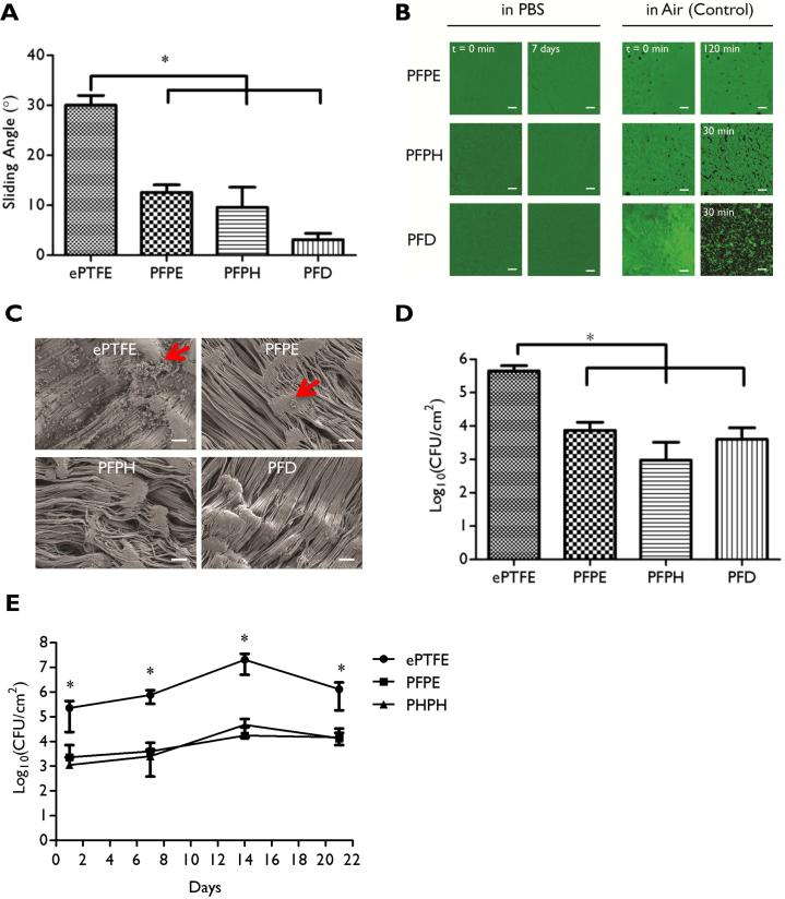
Figure 1. In vitro characterization of ePTFE-SLIPS and anti-bacterial adhesion behavior.
(A) Slippery function of SLIPS was measured by tilting angle assay. (B) Reflection confocal microscopy images of lubricant-infused surfaces submerged over varying intervals in PBS or air. (C) SEM images of ePTFE or ePTFE-SLIPS after two days in culture with S. aureus (Scale bar: 10 μm). Red arrow: bacteria. (D) Colony-forming units (CFU) of ePTFE or ePTFE-SLIPS after exposure to S. aureus for two days. (E) CFU of ePTFE or ePTFE-SLIPS incubated in 50% rat serum for varying intervals and subsequently exposed to S. aureus for 48h. Error bars represent mean ± s.d. from at least 3 replicates, *p < 0.05, ePTFE vs PFPE, PFPH and PFD.

