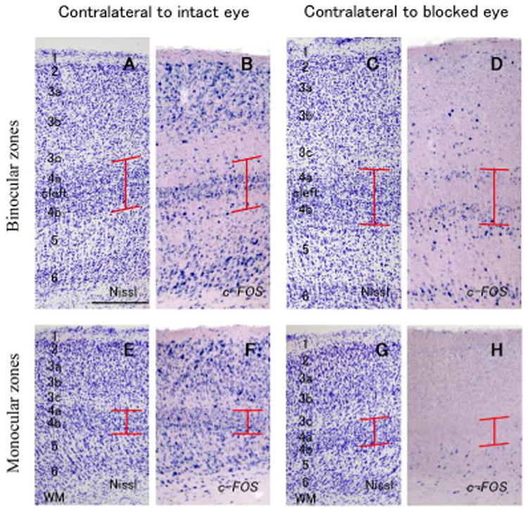
Figure 5.

Higher magnifications of coronal sections of V1 (ID13-54). A-1 D are in binocular zones (dorsal V1) and E-H are in monocular zones (ventral V1). A, B, E and F are contralateral to the intact active eye, and C, D, G and H are contralateral to the blocked eye. A, C, E and G are adjacent sections stained for Nissl substance, and B, D, F and H are stained for c-FOS mRNA. Red lines indicate the extent of layer 4. Scale bar = 250 μm.
