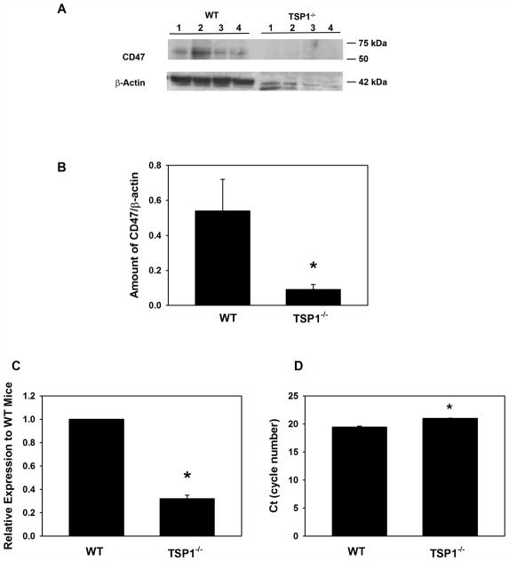
Fig. 5. Amount of CD47 in Lacrimal Glands in TSP1−/− Compared to WT Mice.
Lacrimal glands from female WT and TSP1−/− mice at 12 weeks old were removed, homogenized and proteins subjected to western blot analysis using antibodies against CD47 and β-actin. Western blot is shown in A. Each lane represents an individual animal. Gels were scanned and amount of CD47 (B) was standardized to amount of β-actin. Data are mean ± SEM from 4 individual animals. RNA was isolated from 12 week old LGs of female WT and TSP1−/− mice and cDNA generated. qPCR was performed using primers for CD47. Data are expressed as ratio of cytokines from TSP1−/− mice relative to WT mice from 3 mice and is shown in C. The number of cycles required to detect a fluorescent signal (cycle threshold, Ct) was measured and plotted in D. * indicates significant difference from values of WT mice.

