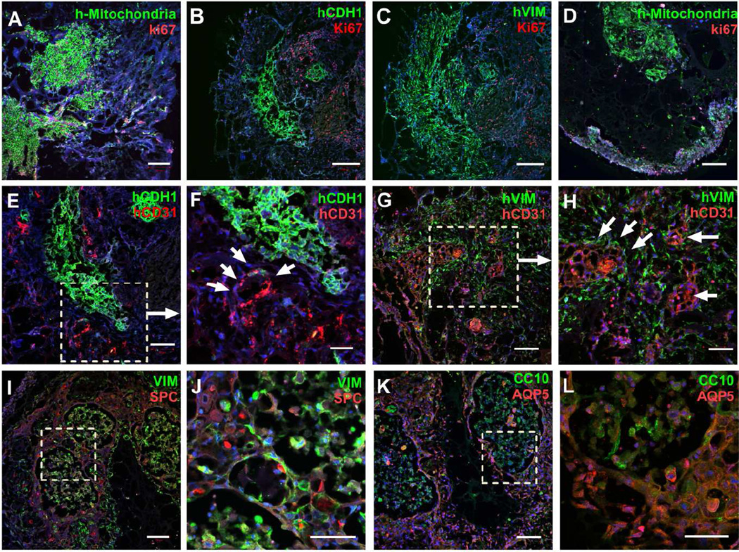
Figure 7.
Ectopic transplantation of human airway organoids. Human mitochondria (green, A, D), human E-Cadherin (green, B) human Vimentin (green, C), and proliferating cells (Ki67, red, A–D) were assessed by immunostaining at the week 1(A–C) and week 6 (D). Human endothelial cells were assessed by human CD31 (red) co-staining with human E-cadherin (green, E, F) and human Vimentin (green, G, H) at week 1. SPC (red) were used for additional epithelial staining with low- and high- power magnification (I, J), as was double staining for AQP5 (red) and CC10 (green) with low- and high- power magnification (K, L). Scale bars, 200µm (B, C), 100µm (A, D, E, G, I, K), 50µm (F, H, J, L). n=4.

