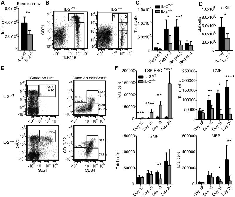
Figure 1. IL-2−/− mice develop bone marrow failure and HSC dysregulation.
Total BM was isolated from 20 day old mice femurs and counted to determine total cellularity (A) and stained for TER119 and CD71 to identify red blood cell developmental stages (B-C). Regions 1-4 correlate with progressive stages of RBC differentiation with region 1 and 4 comprising the least and most mature RBCs, respectively. RBC-lysed BM was analyzed by flow cytometry for the total number of Lin−c-kit+ cells (D). BM was analyzed for the frequency and total number of Lin−Sca1+c-kit+ (LSK) HSCs and Lin−c-Kit+Sca1−CD34+CD16/32− CMPs, Lin−c-Kit+Sca1−CD34+CD16/32+ GMPs, and Lin−c-Kit+Sca1−CD34−CD16/32− MEPs from 12, 16, 18, and 20 day old mice (E-F). Flow plot shows representative data from 20 day old mice (E). (A-E) Data are from at least 2 independent experiments with n=6-10 mice per group. (F) Data are from 1-2 experiments with n=2-8 mice per group. * p < 0.05; ** p < 0.01; *** p < 0.001; **** p < 0.0001 based on students t test.

