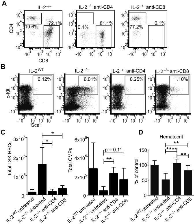
Figure 4. CD4+ and CD8+ cells contribute to bone marrow failure in IL-2−/− mice.
Mice were treated with depleting antibodies to CD4 and CD8α from 8 to 16 days of age and euthanized at day 19. Representative flow plots of bone marrow stained for CD4 and CD8 to determine peripheral depletion efficacy, gated on live CD3+TCRβ+ events (A). Representative flow plots of BM LSK HSCs gated on live Lin− cells (B). Total LSK HSCs and CMPs from BM (C). Hematocrit relative to untreated IL-2WT mice at 19 days (D). Data are from 3 independent experiments with 3-5 mice per group. * p < 0.05; ** p < 0.01 based on students t test.

