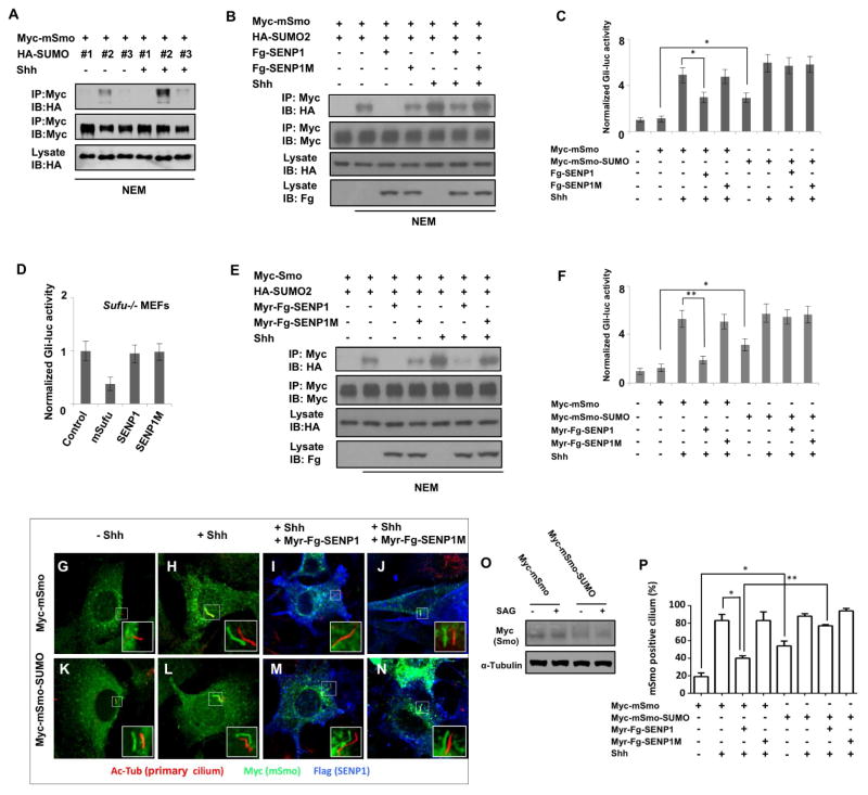
Figure 7. Sumoylation regulates mSmo ciliary accumulation and Shh signaling.
(A) Western blots to detect SUMO conjugated mSmo derived from NIH3T3 cells transfected with Myc-mSmo and HA-tagged SUMO1, SUMO2 or SUMO3 and treated with or without Shh. (B, E) Western blots to detect SUMO-conjugated mSmo derived from NIH3 cells transfected with the indicated constructs and treated with or without Shh. (C, F) Gli-luc reporter assay in NIH3T3 cells transfected with the indicated constructs and treated with or without Shh. Data are means ± SD from three independent experiments. * P<0.05, ** P<0.01. (D) Gli-luc reporter assay in Sufu−/− MEFs transfected with the indicated constructs. (G–N) NIH 3T3 cells stably expressing Myc-mSmo (G–J) or Myc-mSmo-SUMO (K–N) were transfected with or without Myr-Fg-SENP1 or Myr-Fg-SENP1M and treated with or without Shh, followed by immunostaining with antibodies against acetylated tubulin (primary cilium, red), Myc tag (mSmo, green) and Flag tag (SENP1, blue). The insets show the enlarged view of the selected regions with shifted overlays of primary cilium (red) and mSmo (green) immunofluorescence signals. (O) Western blot analysis of cell extracts form Myc-mSmo or Myc-mSmo-SUMO expressing cells treated with or without SAG. (P) Quantification of mSmo positive primary cilia as indicated by the percentage of cells (50 cells were examined for each group) exhibiting ciliary Myc-mSmo signals. Data are means ± SD from two independent experiments. * P<0.05, ** P<0.01.

