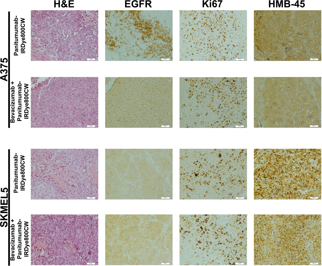
Figure 1.
Histologic assessment of A375 and SKMEL5 tumor specimens 14 days after systemic injection of panitumumab-IRDye800CW or pretreatment with bevacizumab followed by panitumumab-IRDye800CW. Initial staining was with hematoxylin and eosin (H&E). Epidermal growth factor receptor (EGFR) immunohistochemical staining and reactivity analysis was performed. Membrane EGFR reactivity was based on the following scores: 0 = no staining to < 10% of tumor cells staining, 1+ = light and incomplete staining in > 10% of tumor cells, 2+ = moderate and complete staining of > 10% of tumor cells and 3+ = intense and complete staining > 10%. Ki67 staining was done to evaluate to cell viability after therapy. Human melanoma black (HMB-45) anti-melanoma antibody was performed to confirm melanoma tumor cells.

