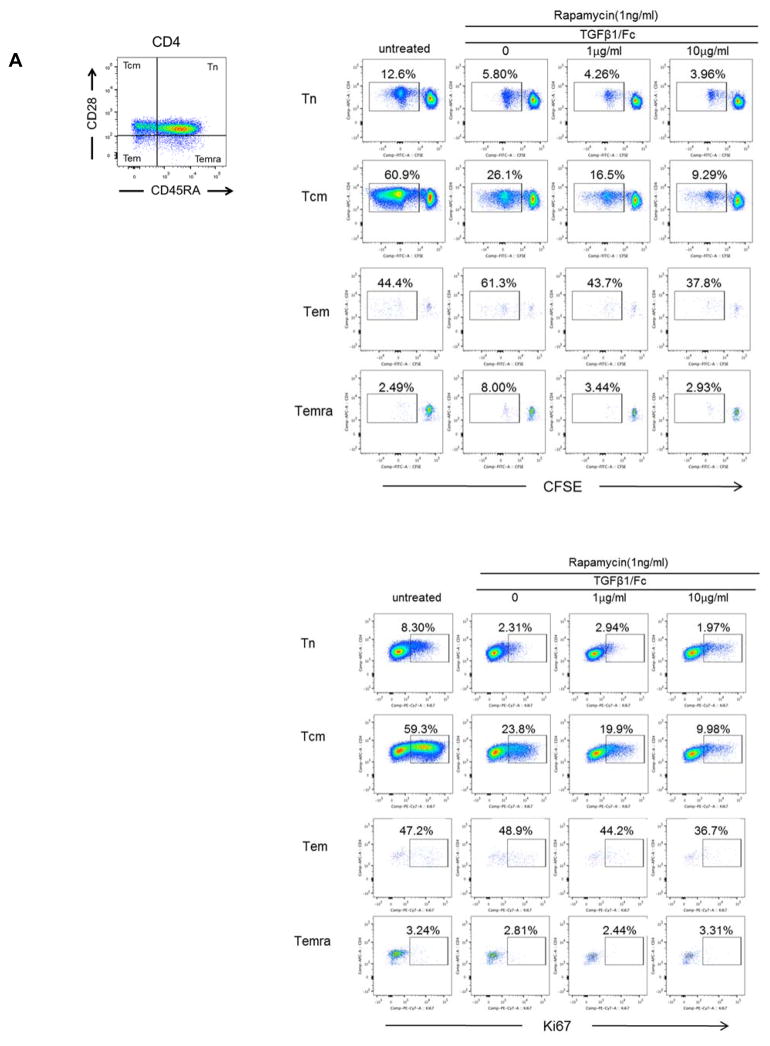
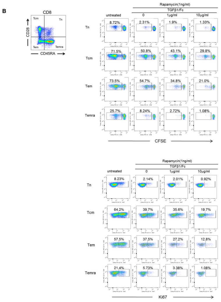
Figure 2. TGFβ1/Fc augments the suppressive effects of rapamycin on memory T cell proliferation.
(A) Percent of proliferation and Ki67 expression by CD4+ naïve and Tmem subsets in response to allostimulation in the presence of rapamycin ± TGFβ1/Fc. (B) Similarly, CD8+ naïve and Tmem subsets were also evaluated for percent of proliferation and Ki67 expression. CFSE-labeled PBMC were cultured for 4 – 5 days with T cell-depleted allogeneic PBMC in the absence or presence of rapamycin ± TGFβ1/Fc. At the end of the culture, T cell subsets were assessed by flow cytometry based on CD45RA and CD28 expression: naïve (Tn)- CD45RA+CD28+, central memory (Tcm)- CD45RA−CD28+, effector memory (Tem)- CD45RA−CD28−, and terminally-differentiated effector memory (Temra)-CD45RA+CD28−. All results are representative data of three independent experiments using different responders and stimulator-responder pairs. PBMC, peripheral blood mononuclear cell.


