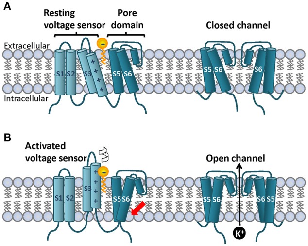
Figure 2.

Topology of a canonical voltage gated ion channel subunit on the lipid bilayer and cartoon representation of the lipoelectric hypothesis mechanism of PUFA-dependent modulation of Shaker-like K+ channels. On the cell membrane four subunits co-assemble to for the ion channel. (A) Each subunit contains six transmembrane segments and N-and C-terminal domains. S1–S4 segments form the voltage sensor and S5–S6 form the ion pore. The S4 segment contains a variable number of positively charged residues known as gating charges that detect small changes on the electric field on the membrane. In response to prolong depolarizations the S4 moves upward perpendicular to the membrane and rotates. (B) This process is called voltage sensor domain activation and it is indicated by the white arrow. Once the four individual S4 are on the upstate the activation gate located at the base intracellular site of the pore domain can open to allow K+ flow (red arrow). A simplified PUFA structure is depicted with the head group located at the extracellular bilayer interface and the bilayer center is depicted in yellow. The PUFA head group interacts electrostatically with the upper gating charges of the S4 promoting mainly channel opening (red arrow). Top and bottom right panels show the front view of the S5 and S6 segments in the closed and open state. Two subunits are depicted for simplicity.
