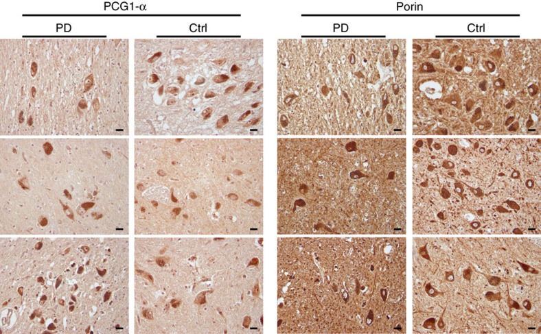
Figure 5. Immunohistochemistry for porin and PGC-1α in dopaminergic substantia nigra neurons of individuals with PD and controls.
Representative sections are shown from three individuals with PD and three controls (Ctrl). There is no detectable difference in staining intensity or distribution between individuals with PD and controls. All pictures have been taken at × 400 magnification. Scale bars, 20 μm.

