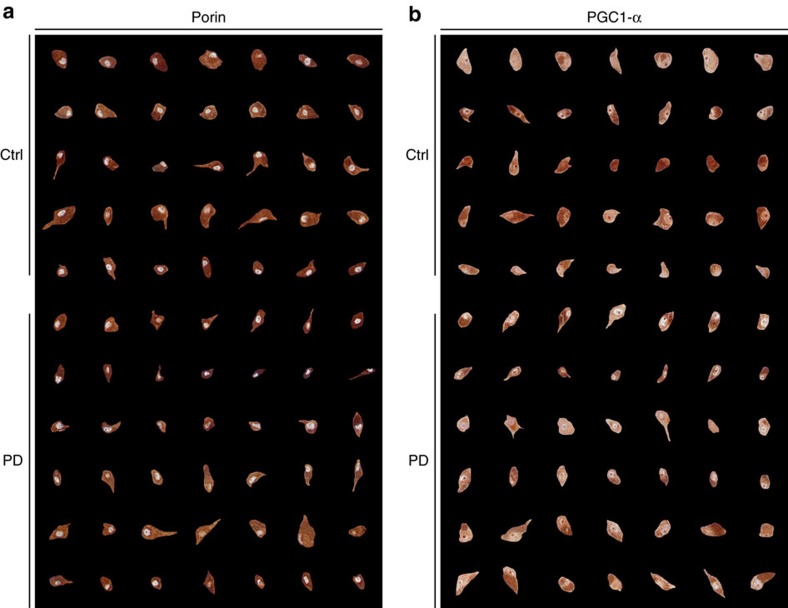
Figure 6. Single-cell montage of porin and PCG-1α-stained dopaminergic substantia nigra neurons of individuals with PD and controls.
The photomontages show representative examples of individual dopaminergic neurons from the ventrolateral tier of the substantia nigra pars compacta, stained with antibodies against porin (a) and PGC-1α (b). Each row shows neurons from the same individual. In both panels, the top five rows are controls and bottom six rows individuals with PD. Magnification: × 400.

