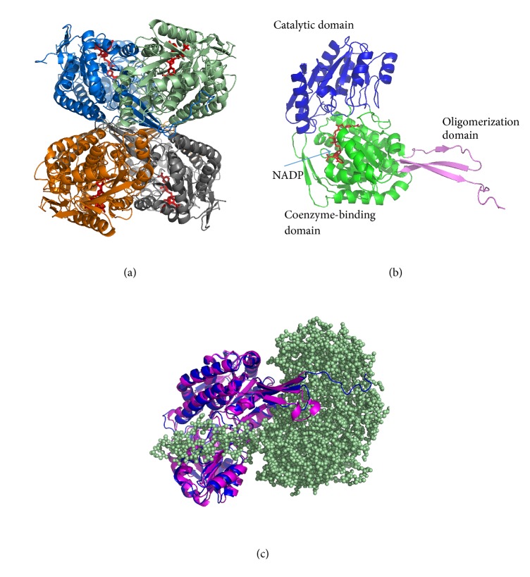
Figure 3.
Overall structure of AlDHPyr1147. The coenzyme is in red. (a) The structure of the tetramer. The subunits that form the tetramer are in blue, grey, brown, and pale green. (b) The structure of the subunit. The catalytic, coenzyme-binding, and oligomerization domains are in blue, green, and magenta, respectively. (c) The superposition of the subunit of aldehyde dehydrogenase from Streptococcus mutants, PDB entry 2EUH (magenta), and the subunit of AlDHPyr1147 (blue). The second subunit of AlDHPyr1147 that forms a dimer with the first subunit is shown as pale green spheres.

