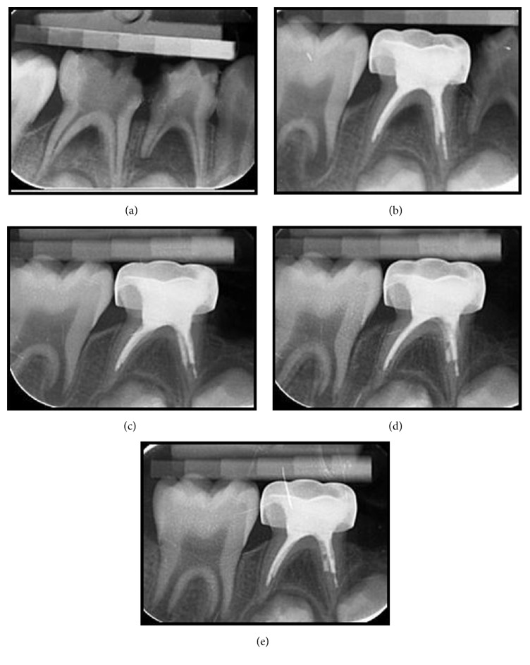
Figure 3.
(a) Radiograph of second primary mandibular molar with a lesion in the interradicular area in MTA group. (b) Radiograph of the tooth after treatment. (c) Radiograph of the tooth at the 3rd month visit. (d) Radiograph of the tooth at the 12th month visit. (e) Radiograph of the tooth at the 18th month visit.

