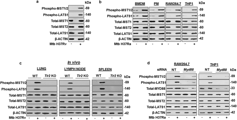
Figure 1. Mycobacterium tuberculosis (Mtb)-driven TLR2 signaling activates Hippo pathway.
(a) Mouse primary peritoneal macrophages were infected with Mtb H37Rv for 1 h to assess the phosphorylated forms of MST1/2, LATS1 and the total levels of MST1, MST2 and LATS1 by immunoblotting. (b) Hippo pathway activation was assessed by immunoblotting in BMDM, PM, RAW264.7 and THP1 macrophages infected with Mtb H37Ra for 1 h. (c) Hippo pathway was assessed in lung, lymph node and spleen of WT and Tlr2 KO mice infected with Mtb H37Rv by aerosol route. (d) Myd88 was knocked down in RAW264.7 and THP1 macrophages followed by infection with Mtb H37Ra for 1 h to check the status of Hippo pathway by immunoblotting. β-ACTIN was used as loading control for immunoblotting experiments on total cell lysates. Blots are representatives of three independent experiments. siRNA, small interfering RNA; WT, wild type; NT, non-targeting; BMDM, bone marrow derived macrophages; PM, peritoneal macrophages; THP1, human leukemia monocytic cell line; h, hour; KO, knock out.

