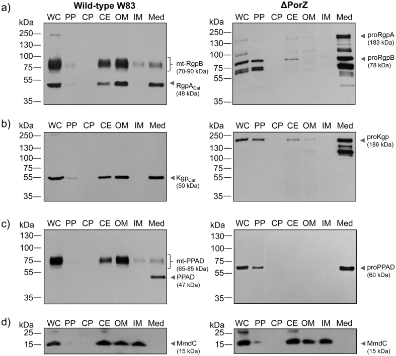
Figure 2. Subcellular location of gingipains and PPAD.
Whole cells (WC) of wild-type (W83; left panel) and ΔPorZ (right panel) P. gingivalis strains were proportionately fractionated into periplasm (PP), cytoplasm (CP), cell envelope (CE), outer membrane (OM), inner membrane (IM) and culture medium fractions (Med; 10-fold concentrated); and probed for (a) Rgps, (b) Kgp, (c) PPAD by Western blotting with specific monoclonal antibodies and (d) biotinylated IM protein (MmdC) through reaction with streptavidin conjugated to horseradish peroxidase. The pinpointed and labeled bands correspond to: (a) catalytic domain of RgpA (RgpAcat) and membrane-type RgpB (mt-RgpB) in the wild type (left panel) and unprocessed pro-RgpA and pro-RgpB in ΔPorZ (right panel); (b) catalytic domain of Kgp (Kgpcat) in the wild type (left panel) and unprocessed pro-Kgp in ΔPorZ (right panel); (c) mature PPAD and membrane-type PPAD (mt-PPAD) in the wild type (left panel) and unprocessed pro-PPAD in ΔPorZ (right panel); and (d) MmdC in the wild-type (left panel) and ΔPorZ (right panel) strains.

