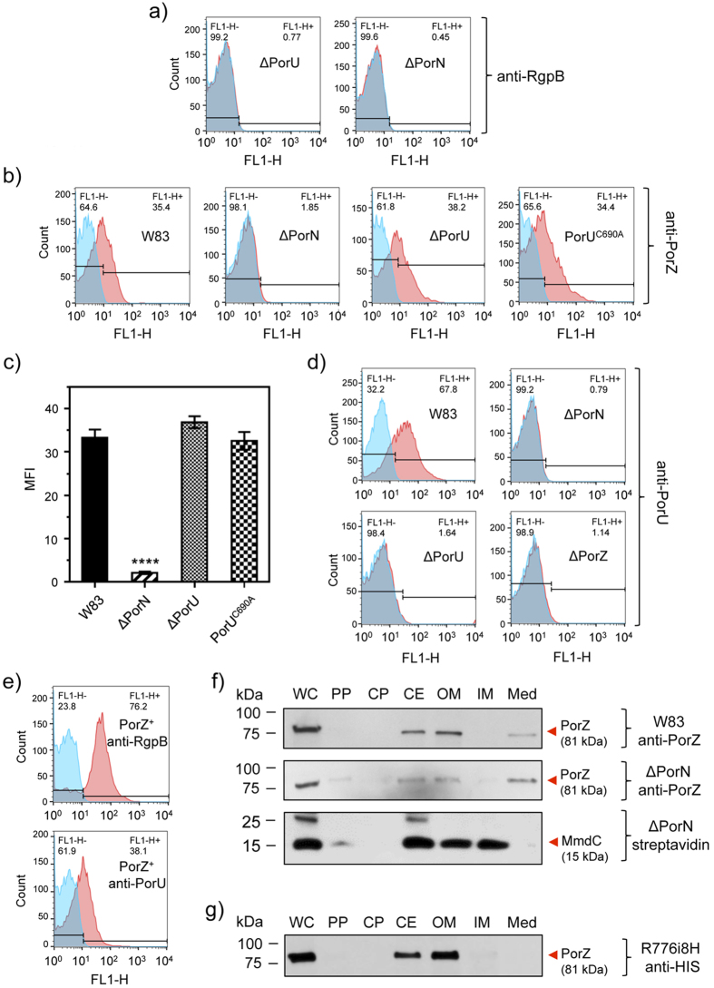
Figure 7. PorZ is secreted via T9SS with an intact CTD independently of PorU.
Flow cytometry analysis using (a) anti-RgpB mAbs in ΔPorU and ΔPorN; (b) anti-PorZ antibodies in the wild type (W83), PorN-null mutant (ΔPorN), PorU-null mutant (ΔPorU), and PorU active-site inactivation mutant (PorUC690A); (c) PorZ surface exposure as mean fluorescent intensity (MFI) in different strains calculated from flow cytometry analysis (in duplicates) from three different cultures. Significant differences between the wild type and mutants were analyzed by one-way ANOVA with Bonferroni’s correction; ****P < 0.0001. Flow cytometry analysis using (d) anti-PorU antibodies in the wild type (W83), PorN-null mutant (ΔPorN), PorU-null mutant (ΔPorU), and ΔPorZ; (e) anti-RgpB and anti-PorU in porZ complemented strain (PorZ+). The result of using specific antibodies (red surface) and the negative isotype control (blue surface) are shown. (f) Western blot analysis of subcellular locations of PorZ in the ΔPorN mutant by probing with anti-PorZ antibodies as compared to the wild type (W83). Streptavidin conjugated to horseradish peroxidase was used to detect MmdC, a biotinylated IM-associated control protein. (g) Same as (f) but using anti-His antibodies to detect the CTD of PorZ in the strain expressing PorZ with an octahistidine tag at the C-terminus (R776i8H). Bacterial cultures were fractionated as described in the Methods section.

