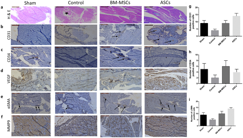
Figure 1. Representative histological analysis of hind limb muscles: Gastrocnemius muscles were collected after 4 weeks of cell therapy.
Tissue samples were stained with: (a) H & E showing muscle degeneration in the ischemic control group and infiltration of lymphocytes (*) compared to normal looking muscles in the BM-MSCs and ASCs treated groups (b) Positive staining for-CD31, in transplanted mice, especially In the ASCs-transplanted group (c) CD34 expression is pronounced in the BM-MSC-transplanted group (d) Increased expression of VEGF especially in the ASC-treated group (e) Staining with anti-αSMA is more pronounced in the ASCs group (f) staining of both tissues with anti-MMP9. Quantitative evaluation of the expression levels of CD31 (g), CD34 (h) and αSMA (i) was evaluated by counting the number of positive cells in each group. Data are shown as mean ± S.D. (error bars). Scale bars, 200 μm.

