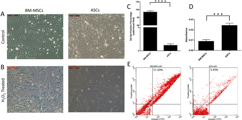
Figure 2. ASCs are more resistant to oxidative stress-induced senescence than BM-MSCs: BM-MSCs and ASCs were exposed to oxidative stress by treating cells with a dose of 600 μM H2O2.
(A) Control cells, (B) H2O2 treated cells showing more than 90% of BM-MSCs positive for SA-β-gal, and ASCs negative for SA-β-gal. (C) Representative images are displayed and data are shown as mean ± S.D. (error bars) of counted SA-Gal positive cells from 5 microscopic fields of 4 independent replicates. *****p < 0.0001. Scale bars, 200 μm. (D) MTT assay (5 mg/ml) to evaluate the proliferation rate of BM-MSCs and ASCs after H2O2 treatment. Formazan absorbance at 570 nm with reference to 630 nm expressed as a measure of cell proliferation [***p < 0.05]. (E) Cellular apoptosis after H2O2 treatment was measured by FACS analysis using Annexin-V and PI staining.

