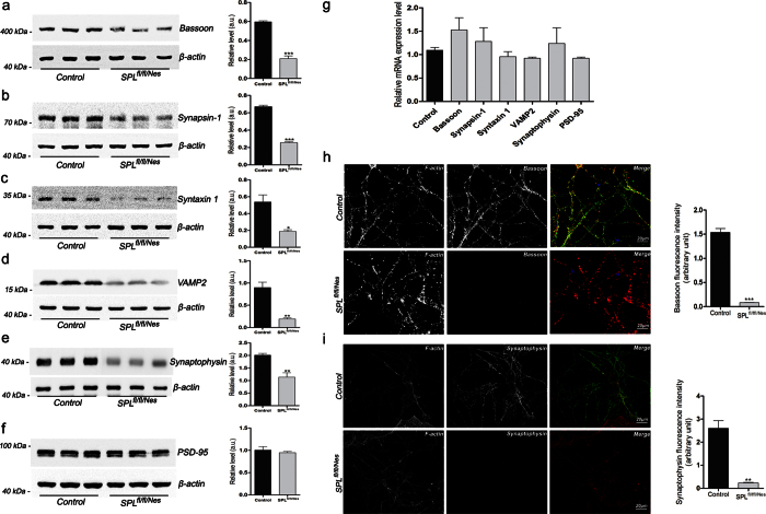
Figure 4. Expression of presynaptic proteins is reduced in SPLfl/fl/Nes mice.
(a–f) Protein amounts were assessed by immunoblotting (unpaired t-test, PBassoon = 0.0002, PSynapsin-1 < 0.0001, Psyntaxin 1 = 0.0157, PVAMP2 = 0.0048, PSynaptophysin = 0.0099), and (g) transcript amounts were determined by qRT-PCR in hippocampi of 6-month-old mice. Immunostaining of the presynaptic marker proteins (h) Bassoon and (i) synaptophysin in cerebellar granule cells after 2 weeks in culture (unpaired t-test, PBassoon < 0.0001, PSynaptophysin = 0.0034). F-actin (red), bassoon and synaptophysin (green).

