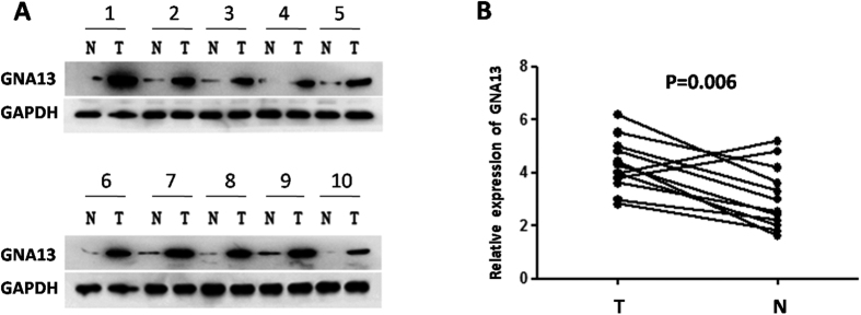
Figure 1. The expression of GNA13 in HCC by Western blotting and qPCR.
(A) Among 12 HCC cases, increased expression of GNA13 was detected via western blotting in 10 pairs of HCC tissues compared with the matched non-cancerous liver tissues. The expression levels were normalised to those of GAPDH. (B) The mRNA expression of GNA13 was significantly upregulated in 10/12 pairs of HCC tissues based on qPCR. The expression levels were normalised to those of GAPDH. N, adjacent normal liver tissue; T, HCC tissue.

