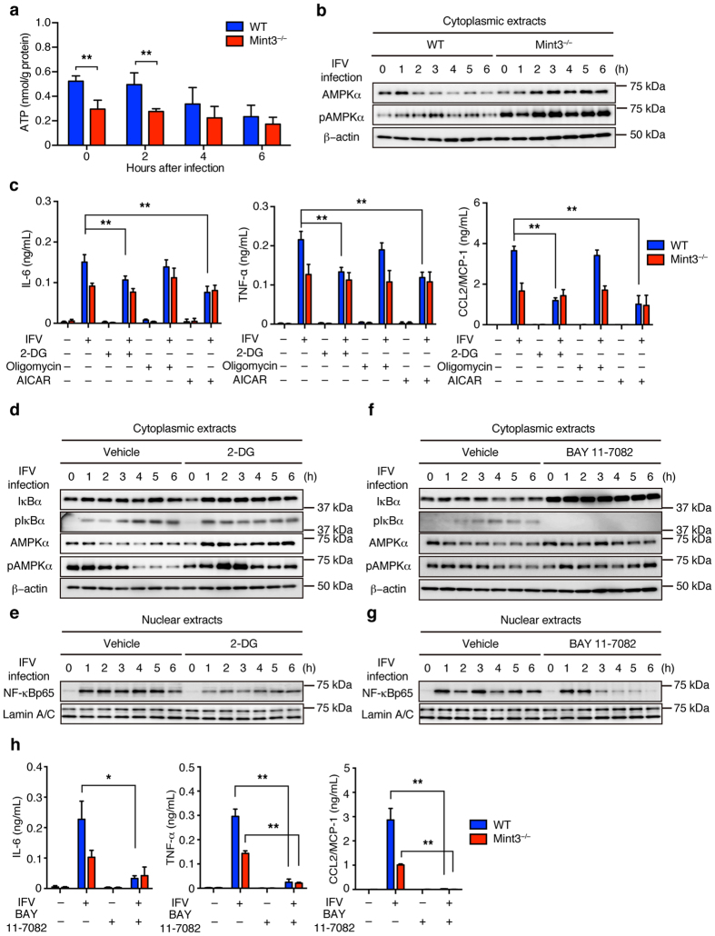
Figure 5. Loss of Mint3 affects NF-κB signalling via both AMPK activation and IκB accumulation in BMMFs.
(a) ATP content of BMMFs after IFV infection. (b) AMPK expression in cellular extracts of WT or Mint3−/− BMMFs after IFV infection. (c) BMMFs were pretreated for 1 h with control vehicle (−), 2-DG (100 μg/mL), oligomycin (5 μg/mL), or AICAR (1 mM) and then infected with 106 PFU (M.O.I = 10) IFV for 24 h. IL-6, TNF-α, and CCL2/MCP-1 production in the cell culture supernatants was measured by ELISA. (d,e) IκBα and AMPK expression in cytoplasmic extracts (d) and NF-κB expression in nuclear extracts (e) of WT or 2-DG-treated WT BMMFs after stimulation with IFV. (f,g) IκBα and AMPK expression in cytoplasmic extracts (f) and NF-κB expression in nuclear extracts (g) of WT or BAY 11-7082-treated WT BMMFs after stimulation with IFV. (h) BMMFs were pretreated for 1 h with control vehicle (−) or BAY 11-7082 (15 μM) and then infected with 106 PFU (M.O.I = 10) IFV for 24 h. IL-6, TNF-α, and CCL2/MCP-1 production in the cell culture supernatants was measured by ELISA. Data are presented as the means ± SD of triplicates and are representative of two independent experiments. **P < 0.05, **P < 0.01 by the Student’s t-test. Unprocessed original scans of blots are shown in Supplementary Fig. S3.

