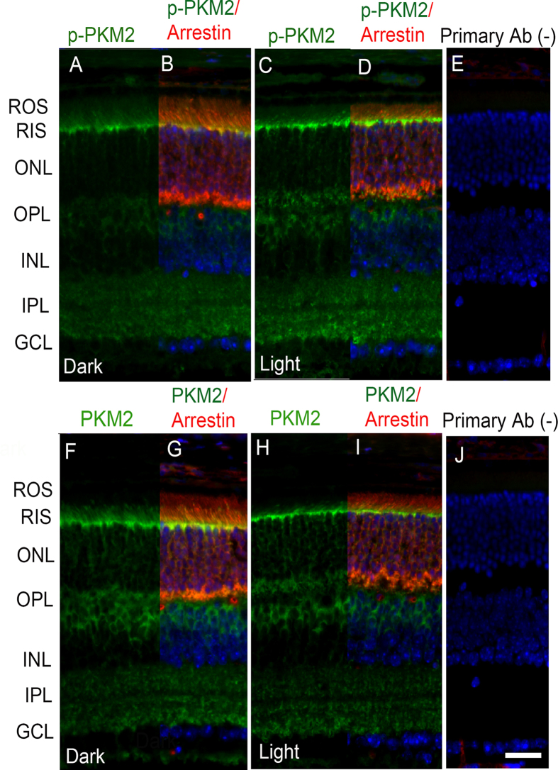
Figure 4. Rhodopsin activation regulates the phosphorylation of PKM2.
Prefer-fixed sections of dark- (A,B,F,G) and light-adapted (C,D,H,I) Rpe65−/− mouse retinas were subjected to immunofluorescence with anti-pPKM2 (A–D), anti-PKM2 (F–I), and arrestin (B,D,G,I) antibodies. Panels (E,J) represent the omission of primary antibody. ROS, rod outer segments; RIS, rod inner segments; ONL, outer nuclear layer; OPL, outer plexiform layer; INL, inner nuclear layer; IPL, inner plexiform layer; GCL, ganglion cell layer. Scale bar 50 μm.

