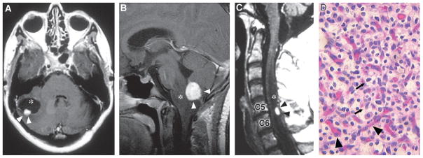
Fig. 10.3.
(A) Axial T1-weighted postcontrast magnetic resonance image (MRI) of a cerebellar hemangioblastoma (arrowheads) with an associated cyst (asterisk) in a 40-year-old woman. (B) Sagittal T1-weighted postcontrast MRI of medullary hemangio-blastoma (arrowheads) with associated brainstem edema (asterisk) in a 12-year-old girl. (C) Sagittal T1-weighted postcontrast MRI of the cervical spinal cord of a 50-year-old man. Hemangioblastoma (black arrowheads) is located in the dorsal spinal cord at C5 and C6, and is associated with a large syrinx (asterix). (D) Hematoxylin and eosin staining of a hemangioblastoma showing the lipid-laden stromal cells (arrows) distributed within a capillary network (arrowheads). (Adapted from Lonser et al., 2003a.)

