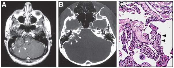
Fig. 10.5.
(A) Axial T1-weighted postcontrast magnetic resonance image shows large heterogeneously enhancing tumor in the right mastoid region (arrowheads). (B) Axial computed tomography through the same region showing the bony erosion of posterior petrous region that often occurs in these tumors (arrowheads). (C) Hematoxylin and eosin stained section showing the typical histologic features of neoplasm, including cuboidal epithelium (arrowheads) in a papillary pattern. (Adapted from Lonser et al., 2003a.)

