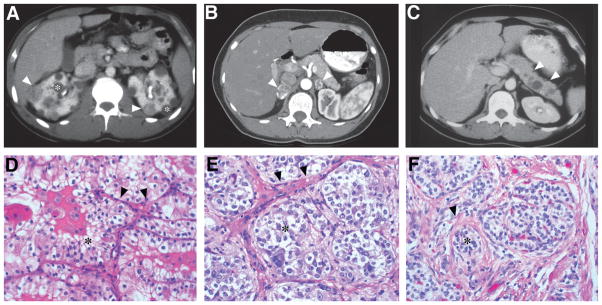
Fig. 10.6.
(A) Bilateral multifocal renal cell carcinoma with both solid (arrowheads) and cystic (asterisk) disease in 22-year-old man. (B) Bilateral pheochromocytomas (arrowheads) with rim enhancement in the adrenal glands of a 29-year-old woman. (C) Pancreatic neuroendocrine tumor (arrowheads) in the body of the pancreas of a 26-year-old woman. (D) Renal cell carcinoma of the clear cell subtype (asterisk) with acinar and tubular architecture embedded in fibrovascular stroma (arrowheads). (E) Pheochromocytomas show a similar appearance and are composed of chromaffin cells. The tumor cells are arranged in rounded clusters (asterisk), separated by endothelial-lined spaces, and have vesicles containing norepinephrine and epinephrine. (F) Pancreatic neuroendocrine tumors show trabecular architecture, small nuclei, and abundant eosinophilic cytoplasm. Nests of tumor cells (asterisk) show focal nuclear atypia with surrounding stromal collagen bands (arrowhead). (Adapted from Lonser et al., 2003a.)

