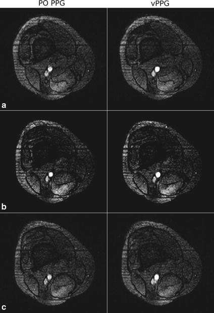
Fig. 13.

Acquired MRA images of an axial slice obtained during PO and vPPG triggering. In a the image acquired using vPPG triggering has a sharply defined vessel compared to the blurred vessel obtained by PO triggering. In b the vessel has a sharp outline and high contrast in both images. In c the image acquired using vPPG exhibits a blurred vessel signal compared to the sharply defined outline that was obtained by PO triggering
