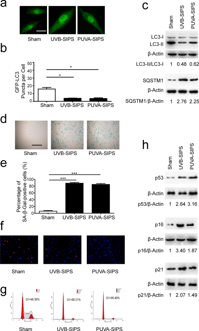
Figure 1. Autophagy is down-regulated in PUVA-SIPS and UVB-SIPS fibroblasts.

a. Cells were transiently transfected with GFP-LC3, and then treated with 10 J/cm2 of PUVA for 14 days or 25mJ/cm2 of UVB twice a day for 5 days to establish PUVA- and UVB-SIPS models. Representative images were taken by confocal microscopy. Scale bars = 50μm. b. The percentage of cells with greater than 10 GFP-LC3 puncta was counted on the images. (means ± SEM of the independent experiments, n = 3, *p < 0.05). c., h. Cells were collected for western-blotting analysis using LC3-, p62-, p53, p16 or p21-specific antibodies. Actin was used as a loading control. The LC3-II/LC3-I, SQSTM1/Actin, p53/Actin, p16/Actin, and p21/Actin densitometric ratios were marked (3 independent experiments gave similar results. See Figure S1). d. SA-β-Gal staining was performed. Scale bars = 100μm. e. The cellular senescence was determined by SA-β-gal staining. Premature senescence cells stained blue. The percentage of SA-β-Gal positive cells was calculated. (means ±SEM of the independent experiments, n = 3, ***p < 0.001). f. The cells were stained by EdU and Hoechst two days post UV-irradiation. g. G1 arrest was analyzed by flow cytometry.
