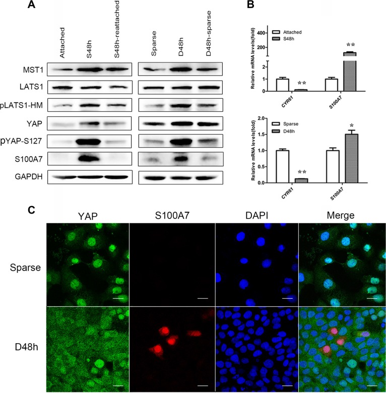
Figure 1. Cell morphology and density regulate S100A7 induction and YAP activity, and subcellular location of S100A7 and YAP are detected in dense cells.
(A) Western blot analyses of S100A7, YAP and pYAP (S127) in the indicated cells. Cells were cultured in suspension for two days (S48 h) and then reattachment for one day (S48 h-reattached). Cells were cultured densely for two days (D48 h) and then relief from dense culture (D48 h-sparse). GAPDH was used as a loading control. (B) The expression of S100A7 and CTGF or CYR61, two endogenous markers of YAP, were analyzed by qPCR. Error bar, SD of three different experiments.*p < 0.05, **p < 0.01; t-test. (C) Dense culture induced S100A7 expression and caused YAP nuclear exclusion in A431 cells. Cells were cultured in dense for two days. Samples were then stained with anti-S100A7 (Abcam) and anti-YAP (CST) antibodies. DAPI is a nuclear counterstain. Scale bar, 20 μm.

