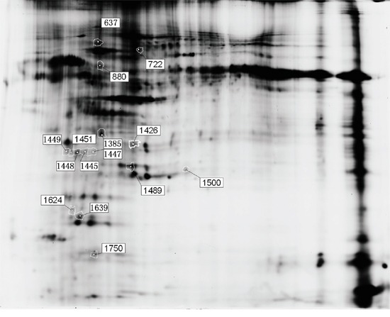
Figure 3. Fifteen differentially expressed protein spots in the two dimensional-fluorescence difference gel electrophoresis (DIGE).

Proteins were extracted from placentas of female SD rats as described in the text. The first dimension was pH 3–10 NL IPG strips, and the second dimension was 12.5% polyacrylamide. The image of fluorescently labeled proteins was acquired using a Typhoon 9400 scanner.
