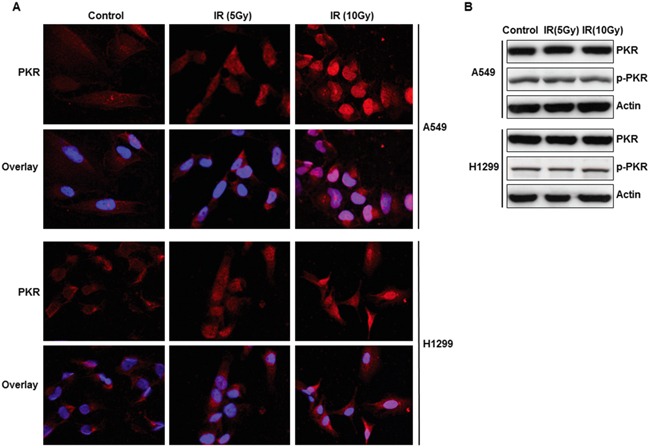
Figure 1. Subcellular localization and expression of PKR in A549 and H1299 cancer cells after radiation treatment.

A. Immunofluorescence microscopy with antibodies against PKR (red) and the nucleus (blue=DAPI staining for DNA in the nucleus) demonstrated accumulation of PKR in the nuclei of A549 and H1299 lung cancer cells after 48 hrs of treatment with radiation. B. Western blot analysis of PKR and p-PKR, protein expression in A549 and H1299 lung cancer cells after 48 hrs of radiation treatment. The expression of actin was used as a control.
