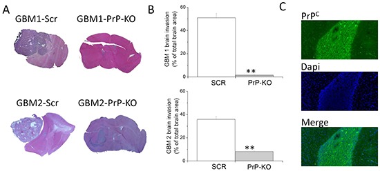
Figure 10. Role of PrPC in the in vivo tumorigenicity of human GBM CSCs.

A. Representative images of tumors developed in NOD/SCID mice orthotopically grafted with GBM1- or GBM2-Scr, and GBM1- or GBM2-PrP-KO cells. After sacrifice of the animals, brains were fixed and stained with hematoxylin and eosin (H&E), showing that GBM1- and GBM2-Scr cells developed larger and more invasive tumors, as compared to the respective GBM PrP-KO cells. B. Quantification of GBM 1 and 2 brain invasion area, evaluated as percentage of total brain area. **p<0.01 vs. respective GBM-Scr cells. C. Representative immunofluorescence analysis of PrP expression (antibody 3F4, green) in tumors originated from GBM1-PrP-KO. Nuclei were counterstained with DAPI (blue).
