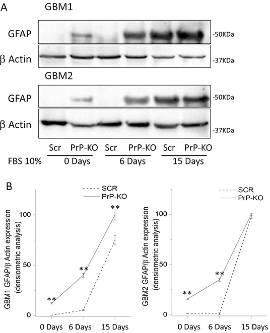
Figure 7.

A. Representative immunoblots of GFAP expression in GBM-Scr and GBM-PrP-KO cells. Cells were analyzed after growing in stem cell permissive medium (time 0), or after cell differentiation, induced by incubation for 6 or 15 days in medium deprived of growth factors and additioned with 10% FBS. Immunoblot analysis of β-actin was performed to verify the total protein input in the cell lysates. B. GFAP expression is reported as densitometric analysis of the gels derived from three independent experiments and expressed as percentage of the highest intensity value of GFAP/β-actin ratio. Cells were analyzed after growing the cells in stem cell permissive medium (day 0), or after inducing cell differentiation by incubation for 6 or 15 days in medium deprived of growth factors and additioned with 10% FBS. **p<0.01 vs. respective GBM-Scr cells.
