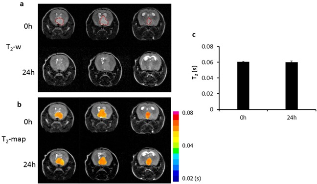
Figure 5. In vivo MRI of the control probe, Aur-L-IO/DiR.

a. To establish the glioma-targeting specificity of the PS-L-IO/DiR probe, the control probe Aur-L-IO/DiR (2.5 mg Fe/kg) was injected into a U87 glioma-bearing mouse. T2-weighted images of the mouse brain acquired before and 24 h post injection revealed no obvious signal change in the tumor region (outlined) at 24 h. T2 maps of the tumor b. and quantitative T2 data c. showed minimal change in T2 at 24 h (mean =60 ± 2) versus the baseline 0 h (61 ± 1 ms).
