
Figure 5. OVA specific CD8+ T cell induction in B16 tumours by exosomes is independent of MHC molecules on exosomes.
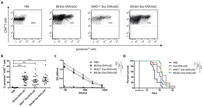
B6 mice were injected s.c. with 200,000 B16/OVA melanoma cells, tumour growth was monitored and mice were treated i.v. with 40 μg B6 Exo-OVA/αGC, MHCI−/− Exo-OVA/αGC or BALB/c Exo-OVA/αGC 4 days after tumour injection. Mice were sacrificed when the tumour reached a volume of 1,000 mm3. A. Representative flow cytometry plots (presented as CD45+, B220−, TCRb+, CD8+, pentamer+ cells) and B. analysis of OVA specific CD8+ T cells infiltrated into the tumour of all four treatment groups. Dots represent a single mouse and data are presented as mean ± SEM. Data were analysed by Kruskal-Wallis with Dunn's multiple comparisons C. OVA-specific IgG antibodies in the sera of the sacrificed animals was determined by ELISA, D. Kaplan-Meier survival curve, data were analysed by Mantel-Cox test. Data represent 2 independent experiments, n=14-15, * P < 0.05, ** P < 0.01, *** P < 0.001.