FIGURE 2.
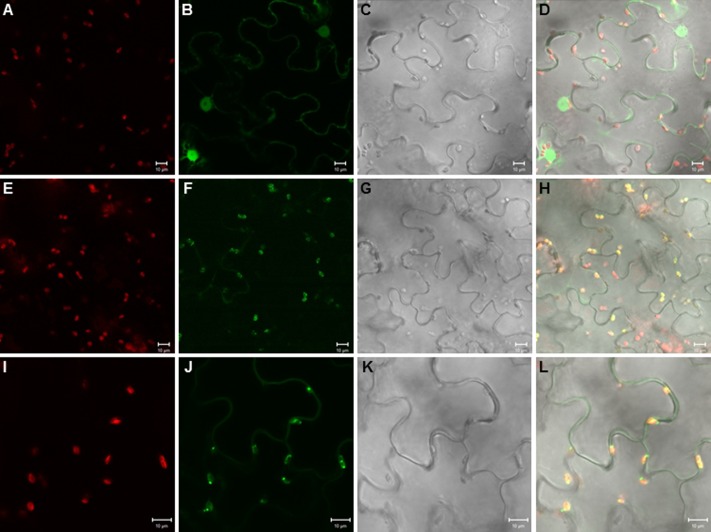
Plastidial localization of the TPS20c protein. Images of tobacco epidermal cells transiently expressing eGFP (A–D), eGFP fused to the TPS20c full length protein (E–H), and eGFP fused to the TPS20c N-terminal transit peptide (I–L) under the control of the CaMV 35S promoter. Images (A,E,I), chlorophyll autofluorescence. Images (B,F,J), eGFP fluorescence. Images (C,G,K), light microscopic images. Images (D,H,L), overlay of chlorophyll autofluorescence, eGFP and light microscopic images. Bar = 10 μm.

