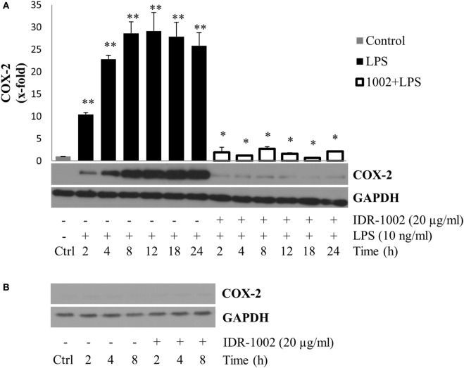
Figure 2.
IDR-1002 inhibited the LPS-induced COX-2 expression in RAW 264.7 macrophages. (A) Macrophages Raw 264.7 were left unstimulated (Ctrl), stimulated with 10 ng/ml of LPS, or pretreated with 20 μg/ml of IDR-1002 for 1 h followed by incubation with 10 ng/ml of LPS for 2–24 h. At the end of each incubation period, COX-2 was analyzed by Western blotting. Protein from Ctrl cells were collected at 4 h. A representative immunoblot of three independent experiments is shown. GAPDH detection was used as control to ensure equal protein loading. (B) At each indicated incubation time, COX-2 expression was measured in untreated control and IDR-1002-treated cells. Values were normalized with GAPDH as protein loading control. This graph is representative of two independent experiments. Data represent means ± SEM (n = 3). **P < 0.05 for LPS compared with the unstimulated control value. *P < 0.05 for 1002 + LPS compared with LPS treatment.

