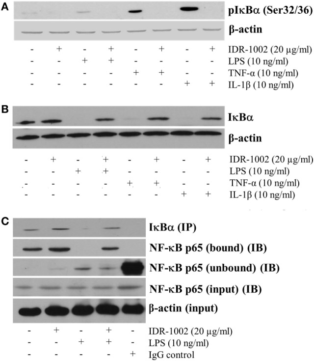
Figure 4.

IDR-1002 inhibited IκBα phosphorylation and degradation in RAW 264.7 macrophages. (A) Macrophages were left unstimulated or pretreated with 20 μg/ml of IDR-1002 for 1 h. Then, macrophages were stimulated for 10 min with either 10 ng/ml of LPS, 10 ng/ml of TNF-α, or 10 ng/ml of IL-1β. Phosphorylated IκBα was detected by Western blot analysis and β-actin was detected to ensure equal protein loading. (B) Macrophages were treated as in (A) except that incubation with LPS, TNF-α, or IL-1β was for 30 min. Total IκBα was detected by Western blot analysis in total cell lysates extracts. β-actin detection was performed to ensure total protein loading. (C) Macrophages were left unstimulated or stimulated with 20 μg/ml of IDR-1002 for 1 h followed by stimulation with 10 ng/ml of LPS for 10 min. IκBα was immunoprecipitated (IP) and eluates were subjected to Western blot to detect NF-κB p65 in the immunoprecipitates of IκBα (bound) or supernatants (unbound) samples (IB). β-actin detection was performed in the input cell extracts to ensure equal protein.
