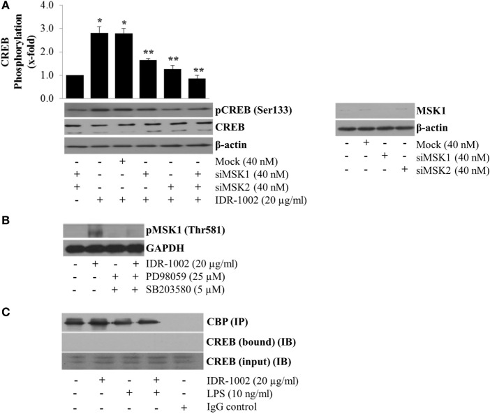
Figure 6.
MSK1-mediated CREB phosphorylation at Ser133 in RAW 264.7 macrophages stimulated with IDR-1002. (A) Macrophages were transfected with non-specific siRNA control (Mock), MSK1 (siMSK1), MSK2 (siMSK2) or MSK1, and MSK2 simultaneously for 48 h. Cells were then stimulated with 20 μg/ml of IDR1002 for 15 min. CREB phosphorylation was analyzed by Western blotting. Detection of β-actin and unphosphorylated CREB in each sample was performed to ensure equal protein loading. The right panel shows detection of MSK1 gene expression when macrophages were treated with MSK1-specific siRNA. In (B), macrophages were left unstimulated (−) or pretreated with 25 μM of PD98059 (PD, an ERK1/2 inhibitor) and 5 μM of SB203580 (SB, a p38 inhibitor) for 30 min, and then stimulated with IDR-1002 for 15 min. Additional controls with 25 μM of PD and 5 μM of SB alone were also included. Under these conditions, phosphorylation of MSK1 was detected by Western blotting. (C) Macrophages were left unstimulated or stimulated with 20 μg/ml of IDR-1002 followed by stimulation with 10 ng/ml of LPS. Immunoprecipitation was performed as previously described and eluates from CBP immunoprecipitates (IP) were subjected to Western blot analysis with CREB antibodies. Then, we detected CREB immunoprecipitated (bound) with CBP and in the input (IB). Control with isotype IgG was also included. Detection of CBP was performed to show equal protein loading. The blots are representative of three independent experiments. Data represent means ± SEM (n = 3). *P < 0.05, compared with the unstimulated control value. **P < 0.05 for siRNA treatment compared with the IDR-1002 value.

