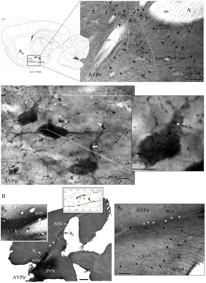
Figure 2.
Anatomo-immunohistochemical features of vasopressin immuno-reactive (AVPir) somata and fibers in the amygdaloidal complex suggest that AVP modulates distinct neuronal circuits. (A) Photomicrograph of AVPir in a sagittal section of rat amygdala at lateral 3.40 mm (Aa, inset: sagittal view of amygdaloid complex, squared region, from the Rat Brain Atlas). Note that the AVP immunopositive fibers (indicated by black arrows) are heterogeneously distributed in the whole amygdala complex and numerous AVP+ cells were inside the bed nucleus of stria terminalis, intra-amygdaloid division (STIA; circumscribed with white dashed line). (Ab) Amplification of a region inside the STIA. Note that there are two types of AVPir+ fibers, regarding their thickness and spatial frequency of varicosities: the type I fibers have thick diameter and frequent varicosities (thick white arrows); the type II fibers are thin and had less frequent varicosities (thin white arrows). Both types are intermingled in the same region. The asterisk indicates the site where one thin-type axon (the axon initial segment was indicated with a hollow arrow, white in both (Ab) and its inset) originated (asterisk) from one proximal dendrite of the respective neuron. (B) A semi-horizontal section of rat brain showing the vicinity of the hypothalamic AVP+ supraoptic (SON) and paraventricular (PVN) nuclei and the massive projection of AVPir fibers toward amygdala and amygdala-hippocampal cortex (AHiCtx; white arrowheads in Ba,Bb). The red line of the atlas inset symbolizes the level and the inclination of the section plan. (Ba) Amplification of the corresponding squared region showing the SON is seen from the semi-horizontal view. (Bb) Amplification of the corresponding squared region of amygdaloid complex. Note the heterogeneous presence of the AVPir fibers in the amygdaloid subdivisions. MeApd, medial amygdala postero-dorsal; CeA, central amygdala; BLA, basolateral amygdala. Scale bars: (A) 500 μm; (Ab) and Inset: 20 μm; (B) 500 μm; (Ba) 100 μm; (Bb) 100 μm.

