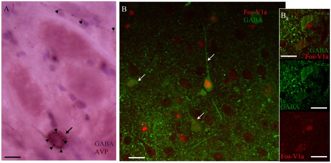
Figure 8.
AVP containing fibers in CeA synapse upon V1a receptor-positive GABAergic neurons activated after EPM. (A) Immunohistochemistry against AVP (arrowheads) and GABA (arrow) showing punctate AVP fibers terminals in close apposition with the GABA-labeled soma. (B) Confocal image of CeA immunoreacted against FOS (red nuclei indicated by arrows); V1a receptor (red punctuated label, indicated by arrowheads) and GABA (green cells). The immunohistochemistry was performed 90 min after the EPM in an animal infused with vasopressin in the CeA. (Ba) A different optical section of the same region showing two GABA+ neurons, one of them negative for V1a receptor showing no expression of Fos, the other one expressing the V1a receptor being Fos+. Scale bars: 10 μm.

