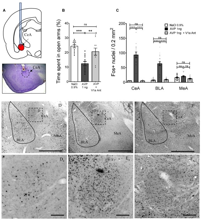
Figure 9.
Altered anxious behavior and Fos expression after AVP infusion into CeA in AFR rats and reversal by co-infusion of V1a antagonist (Manning compound). (A) Upper panel depicts positioning of cannulae in CeA for infusion of vehicle, AVP, or AVP plus V1a antagonist (Manning compound); lower panel is a representative photomicrograph showing the trajectory of the cannula toward the CeA. (B) Bar graph showing the time spent in the open arms of the EPM in animals infused bilaterally with vehicle (NaCl 0.9%), AVP or AVP + V1a antagonist (Manning compound). Infusion of AVP into CeA produced anxiety-like behavior shown as a reduction of the time spent in the open arms (**p < 0.01, ***p < 0.001) compared with vehicle-treated rats. Infusion of AVP + V1a antagonist reversed anxious behavior (decreased time spent in open arms) to control levels (i.e., indistinguishable from vehicle-treated rats). (C) Bar graph showing the quantification of neurons activated (measured by Fos expression) after EPM in the CeA, BLA and MeA in vehicle, AVP, or AVP plus V1a antagonist infused groups. AVP produced a significant increase in the Fos expression in the central (CeA, p < 0.001) and basolateral nuclei (BLA, p < 0.001) that was blunted in the AVP + V1a antagonist group. No effect of the treatments was observed in the MeA. (D–F) Photomicrographs showing the Fos expression patterns seen in the three infusion groups after the EPM, (Da–Fa) are magnifications of the squared regions in (D–F). BLA, basolateral amygdala; CeA, central amygdala; MeA, medial amygdala. Scale Bars: (D–F) 1 mm; (Da–Fa) 0.1 mm.

