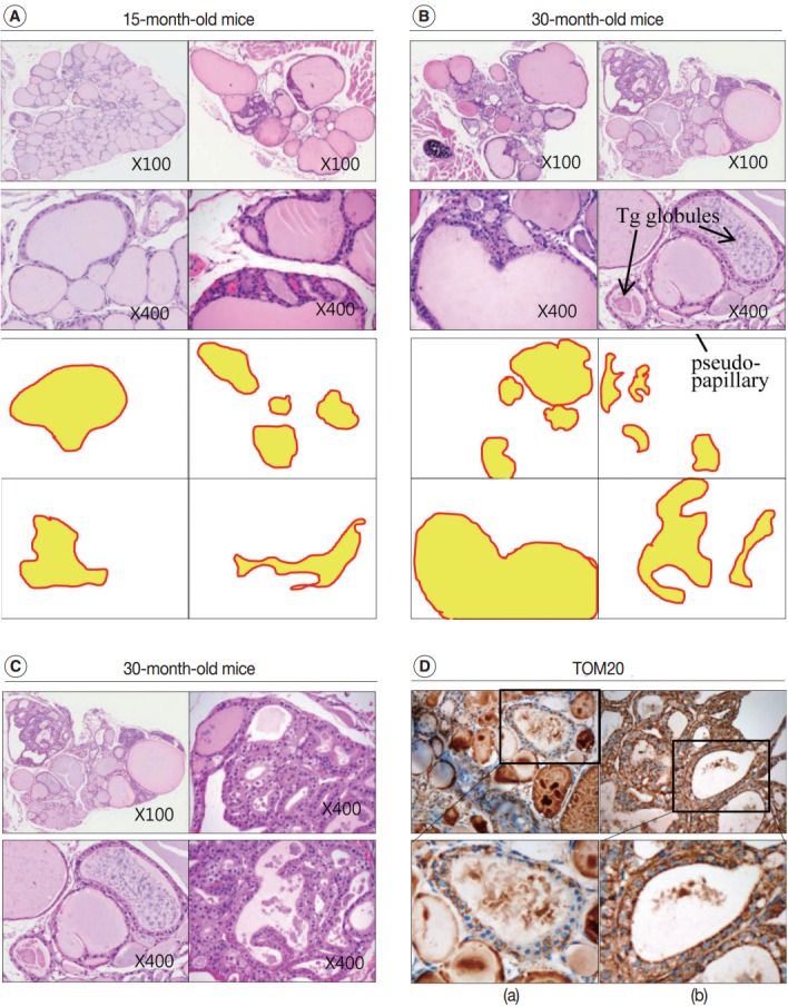
Fig. 3.

Increased irregularity of follicles in the aged mouse thyroid. Hematoxylin and eosin (H&E)–stained sections of thyroid from 15-month-old (A) and 30-month-old (B) mice. (C) Oncocytic change in the follicular cells of aged mouse thyroid stained with H&E. (D) Sections of 30-month-old mouse thyroid stained with anti–translocase of the outer mitochondrial membrane (TOM20) antibody visualized by 3,3'-diaminobenzidine. Non-oncocytic follicular cells are weak for TOM20 (left panels), while oncocytic follicular cells are strongly TOM20-positive (right panels). Tg, thyroglobulin.
