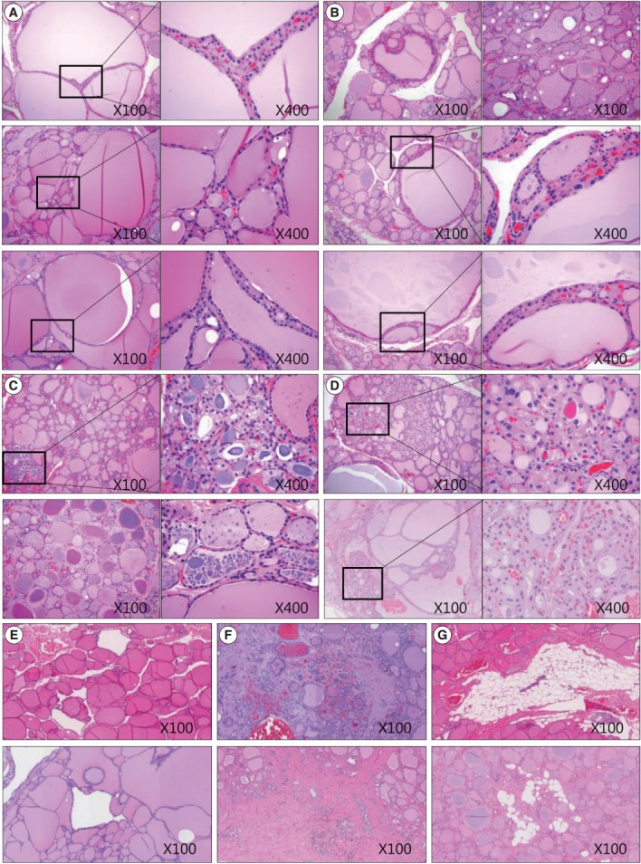
Fig. 5.

Sections of thyroid from elderly females highlighting various morphological changes. (A) Size variation and enlargement of the follicles. (B) Increased irregularity of follicles and Sanderson’s polsters in the wall of large follicles. (C) A large thyroglobulin (Tg) globule (right upper image) and numerous small fragmented Tg globules (right lower image) in the follicular lumen. (D) Oncocytic change in the follicular cells and in those with Sanderson’s polsters. (E) Irregularly dilated follicles lacking colloid. (F) Fibrosis of the extracellular matrix. (G) Fatty infiltration in the extracellular matrix.
