
FIGURE 3.
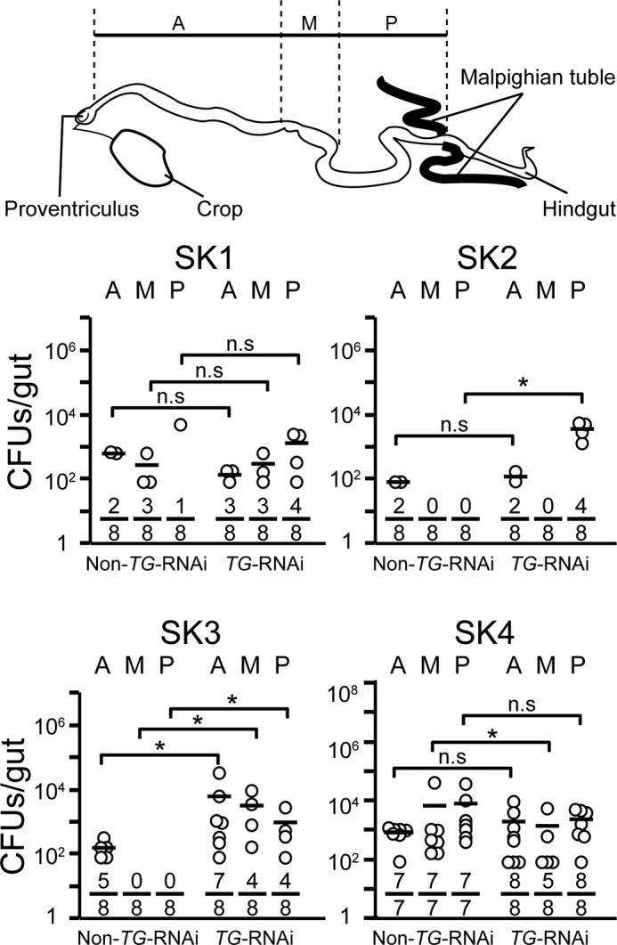
Bacterial colonization ability in fly midguts after transient ingestion of strains SK1–4. The bacterial strains were ingested by 3–5-day-old adult non-TG-RNAi and TG-RNAi flies for 24 h, and then the flies were transferred into sterile vials containing sterile food. After a 5-h incubation, midguts were removed and dissected into three regions: the anterior midgut (A), middle midgut (copper cell region) (M), and posterior midgut (P). The three dissected parts were homogenized and spotted onto the strain-specific agar plates, and the resulting bacterial colonies were counted as described under “Experimental Procedures.” Top, schematic image of the Drosophila midgut. Horizontal bars represent the median cfu/fly for each ingestion group. Each denominator and numerator of the fractions shows the number of all guts used for the experiments and the number of guts with bacteria growing on agarose plates, respectively. p values were calculated by the non-parametric Mann-Whitney U test. *, p < 0.05; n.s, not significant.